





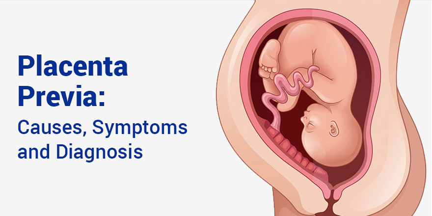
Placenta Previa: Causes, Symptoms and Diagnosis

Placenta previa is amongst the more important complications of the second and third trimesters in obstetrics. The condition is quite unnerving to expectant mothers and families alike. This blog is all about what placenta previa is, types of placenta previa, how it is diagnosed and managed, and how the delivery plan is undertaken if present.
What is Placenta Previa?
The placenta is normally implanted high up in the upper uterine segment, far away from the cervical os. Placenta previa involves an abnormal implantation of the placenta low down in the uterus and it covers all or part of the internal os. The reason why this is a serious issue is that during delivery the cervix needs to dilate, but this cannot happen if the placenta blocks it since the lower segment distends and the cervix effaces. This condition complicates about 0.3-0.5% of pregnancies at term. Detected early, the placenta may shift due to growth of the uterus, while later on special management of placenta previa and delivery are needed.
Types of Placenta Previa
The placenta completely covers the internal os. Vaginal delivery is not possible. Caesarean section is necessary.
Here, the placenta extends to cover the internal os of the cervix partially. Obstetrical management depends on the degree of coverage, and Caesarean section is often required.
The edge of the placenta extends to the margin of the internal os but does not extend beyond it. Depending on the distance, vaginal delivery can be attempted in selective cases after ultrasound measurement.
The edge of the placenta is less than 2 cm from the internal os but does not cover it. There is increased risk associated with this condition compared to low-lying placenta.
What are the Placenta Previa grades?
Placenta previa is categorized according to the proximity of the placenta to the cervix. It is usually categorized in the following ways:
The placenta is located at the lower uterine segment but does not cover the internal cervical os.
The margin of the placenta reaches the internal cervical os but does not obstruct it.
The placenta partially covers the internal cervical os and makes it difficult for vaginal birth to take place.
The placenta entirely covers the internal cervical os and necessitates a Cesarean birth.
Symptoms of Placenta Previa in Pregnancy
Placenta previa signs and symptoms is painless vaginal bleeding. Other placenta previa symptoms include:
Presence of no symptoms does not diminish the clinical importance of this finding.
Placenta Previa Causes & Risk Factors
Some of the common causes and risk factors include the following:
Placenta Previa Diagnosis
Treatment for Placenta Previa
No treatment can reposition the placenta. Placenta Previa Treatments involves ensuring the health of mother and baby until baby is old enough to be delivered safely.
Risks/Complications to Mother and Child
The common complications are:
Delivery Planning with Placenta Previa
Full previa and partial previa are definite indications for a Caesarean section, scheduled in uncomplicated cases at 36-37 weeks; recurrent bleeding necessitates an earlier delivery. The operating surgeon cross-matches blood in advance and expects considerable blood loss during the procedure. If the possibility of accreta spectrum on MRI is present, a multidisciplinary approach is organised pre-operatively involving interventional radiology, urology, and vascular surgery. Prophylactic balloon catheterisation or ureteric stenting can be done prior to the incision by skilled centres.
Does Placenta Previa Resolve by Itself?
During the first trimester of pregnancy, most cases of low placentation turn out to be normal as the uterine body grows. This causes the formation of a larger lower uterine segment, thus resulting in a relatively more elevated location of the placental edge. Full previa identified at the anomaly scan resolves in some patients, while marginal previa found early on has a greater chance of resolution. Placenta previa which persists beyond 32 weeks is highly unlikely to resolve adequately for vaginal delivery.
Also Read: Anterior vs Posterior Placenta: Understanding Placental Position During Pregnancy
Precaution and Lifestyle during pregnancy
For women suffering from placenta previa, some advice includes:
Conclusion
Placenta previa is a condition that should be diagnosed and managed in time to avoid complications for the woman and the child. If you have symptoms like vaginal bleeding without pain during pregnancy, do not ignore them. With today's developments in prenatal care and management of high-risk pregnancy, almost all patients with placenta previa have successful deliveries under proper medical supervision.
If you are planning to get pregnant and need assistance with conceiving, you need to visit the best IVF center in India. Get in touch with a reliable fertility clinic near me to get your fertility treatment from the best IVF doctor in India and the best IVF specialist in India. At Seeds of Innocens IVF, we will support you in your journey towards conception and beyond.
Also Read: Posterior Placenta: Meaning, Types, and Its Impact on Pregnancy
Frequently Asked Questions
1. What is placenta previa, and how common is placenta previa?
Placenta previa is the condition where the placenta attaches itself to the lower uterine segment, obstructing the cervix either partially or wholly. It makes vaginal delivery impossible, poses the risk of haemorrhage, and demands special handling by the obstetrician from diagnosis up to delivery.
2. What are the symptoms of placenta previa?
Painless, bright red vaginal bleeding during the second or third trimester. There will be no tenderness of the uterus, which distinguishes placenta previa from placental abruption. Some patients never have any symptoms, and this condition is detected via routine ultrasound.
3. What causes placenta previa?
It is caused by the improper attachment of the placenta in the lower uterine segment. Uterine scars following Caesarean delivery, D&C, or myomectomy are the predisposing factors that have been identified.
4. How is Placenta Previa diagnosed?
Placenta previa can be diagnosed with the help of transvaginal ultrasonography. Transvaginal ultrasonography is relatively more accurate than a transabdominal sonogram. This disease can be identified through the 18-20 weeks anomaly scan and the 32 and 36 weeks rescan. If placenta accreta is suspected, MRI is done.